Avoiding Vascular Occlusion with Fillers: The 2026 Practitioner’s Safety Guide
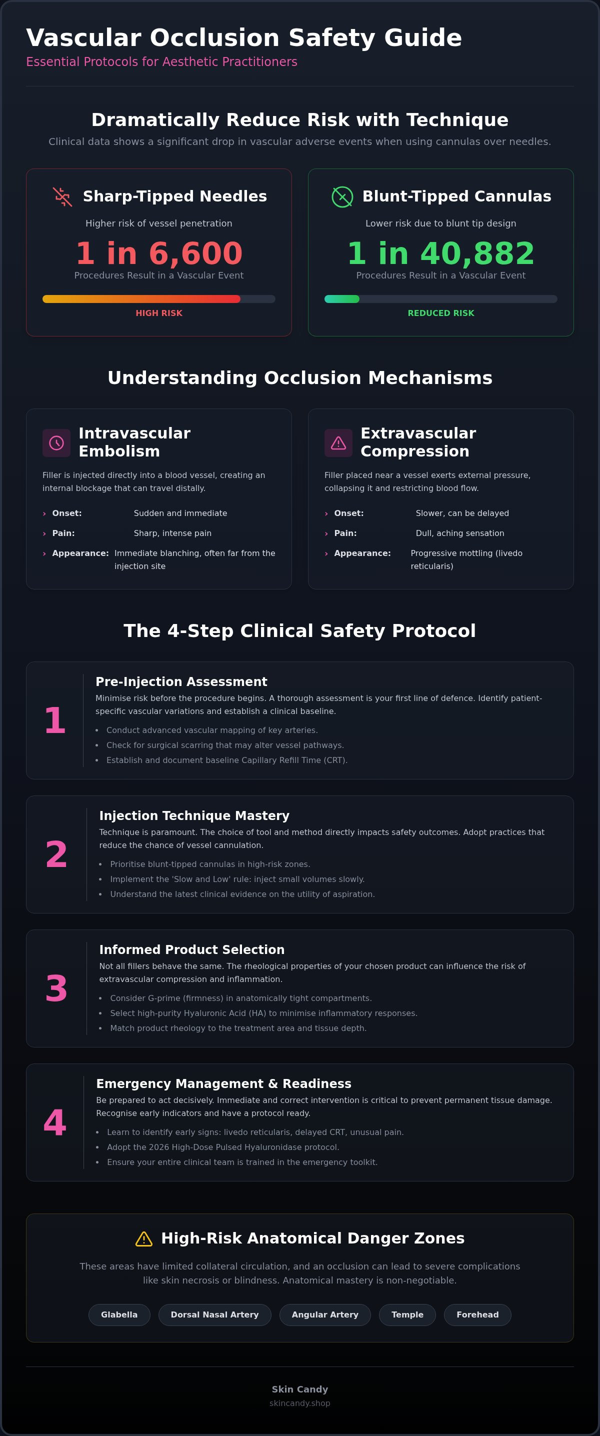
Recent clinical data confirms that vascular adverse events occur in approximately 1 in 6,600 filler procedures, yet this risk drops to 1 in 40,882 when switching from needles to cannulas. For any serious practitioner, avoiding vascular occlusion with fillers is not just a clinical goal; it's the foundation of a sustainable business. You likely feel the constant pressure of navigating high-risk anatomical zones while managing patient expectations for immediate results. The fear of catastrophic complications like skin necrosis is a valid concern that keeps even the most experienced injectors alert.
This 2026 safety guide provides the technical mastery you need to eliminate guesswork. You'll learn to implement the latest clinical protocols and select products with predictable rheological behaviour to minimise vascular risks. We examine the current evidence on aspiration, map out the highest-risk facial zones, and discuss how high-purity HA options like Lumifil and Aesossa support safer outcomes. Master these strategies to maintain a zero-incident record and gain the confidence to handle complex cases with total precision. We provide the logistical certainty and technical insights required to keep your clinic running safely and efficiently.
Key Takeaways
- Master advanced vascular mapping techniques and learn how identifying previous surgical scarring is critical for preventing altered blood flow complications.
- Implement the 'Slow and Low' injection rule and understand the latest clinical data on avoiding vascular occlusion with fillers using blunt-tipped cannulas.
- Discover how rheological properties like G-prime and high-purity HA filler selection influence the risk of inflammatory-induced extravascular compression.
- Learn to identify early clinical indicators of occlusion, such as livedo reticularis and delayed capillary refill time, to ensure immediate intervention.
- Adopt the 2026 High-Dose Pulsed Hyaluronidase protocol as your gold standard for managing adverse events with total clinical confidence.
What is Vascular Occlusion and Why Does it Occur?
Vascular occlusion (VO) is the accidental blockage of a blood vessel by dermal filler material. It represents the most critical safety concern for aesthetic practitioners in 2026. When blood flow is restricted, the affected tissue enters a state of ischaemia. If left untreated, this progresses through a rapid cascade of hypoxia and eventual tissue necrosis. Understanding the underlying mechanisms is essential for avoiding vascular occlusion with fillers and ensuring patient safety.
Clinicians must recognise that injectable filler risks are not uniform across all patients or products. The severity of an occlusion depends on the vessel's diameter, the volume of product injected, and the speed of the injection. In 2026, the focus has shifted from simple anatomy to a more nuanced understanding of how filler rheology interacts with vascular structures.
Intravascular Embolism vs Extravascular Compression
An intravascular embolism occurs when the needle or cannula tip penetrates a vessel wall, allowing the product to enter the lumen directly. This typically causes immediate distal ischaemia, often appearing as sudden blanching far from the injection site. It acts as a mechanical "plug" that stops blood flow instantly. Because the product is inside the vessel, it can also migrate to other areas, increasing the risk of distant complications.
In contrast, extravascular compression happens when the filler is placed outside the vessel but exerts enough pressure to collapse the lumen. This often occurs with high-volume boluses or when using fillers with high G-prime (firmness) in tight anatomical compartments. Practitioners should watch for these differences:
- Embolism: Sudden onset, sharp pain, and immediate blanching.
- Compression: Slower onset, duller ache, and progressive mottling (livedo reticularis).
High-Risk Anatomical Danger Zones
Certain facial areas carry a disproportionate risk of catastrophic complications. The glabella and dorsal nasal artery are frequently cited as high-risk zones. These vessels have limited collateral circulation, meaning a single blockage can lead to permanent skin loss or, in rare cases, blindness via retrograde embolism to the ophthalmic artery.
The angular artery, located near the nasolabial folds, is another high-stakes area. Practitioners must also exercise extreme caution in the temple and forehead. Success here requires a precise understanding of the deep temporal fascia and the exact depth of the vessels. In these zones, the margin for error is non-existent; anatomical mastery is your primary defence against vascular compromise.
Pre-Injection Protocols: Minimising Risk Through Assessment
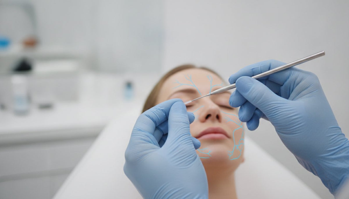
Safety in aesthetics begins long before the syringe is unsealed. For the modern practitioner, avoiding vascular occlusion with fillers relies on a systematic assessment of the patient’s unique vascular architecture. You must conduct thorough mapping to identify the precise path of key arteries, such as the facial and supraorbital vessels. This process is complicated by previous surgical interventions. Scar tissue from rhinoplasty or a facelift can tether vessels in place or force blood flow into collateral pathways. These 'vascular wildcards' increase the risk of accidental penetration because the anatomy no longer follows textbook patterns.
Establishing a clinical baseline is equally vital. Standardise the capillary refill time (CRT) for every patient before you begin. A normal CRT is typically under two seconds. Knowing this baseline allows you to identify subtle changes during the procedure. Document skin temperature and colour amongst all treatment areas to ensure you can spot ischaemic changes early. Maintaining a sterile clinical environment is non-negotiable. Secondary infections can cause delayed swelling that compresses vessels, mimicking or exacerbating a late-onset occlusion.
Patient History and Vascular Integrity
Screen every patient for chronic conditions that compromise vascular health. Smoking and diabetes significantly reduce tissue resilience and slow the healing process. Reviewing FDA guidance on dermal fillers helps in identifying patients with contraindications or higher risk profiles. Always document any history of 'ischaemic events' or previous filler complications. This data informs your choice of product and technique, ensuring you don't repeat past errors. For practitioners who prioritise safety, maintaining a reliable supply of emergency consumables through professional wholesale channels is a fundamental part of risk management.
The Role of Ultrasound in 2026 Aesthetics
In 2026, handheld Doppler and ultrasound devices have moved from optional extras to essential clinical tools. These devices allow you to visualise variant anatomy in real-time. You can map the exact depth and diameter of vessels in high-risk zones like the temples or the nose. Integrating this technology into your just-in-time inventory strategy ensures you have the right tools and fillers ready for every specific anatomical challenge. This proactive approach is the most effective method for avoiding vascular occlusion with fillers while maintaining a high-volume clinical schedule. Using ultrasound to confirm the absence of vessels in your intended injection plane provides an unparalleled layer of safety for both practitioner and patient.
Injection Techniques: Cannula vs Needle and the Aspiration Debate
The choice of delivery tool is a primary factor in avoiding vascular occlusion with fillers. Whilst needles offer precision for supraperiosteal placement, they are inherently sharp. This increases the risk of vessel wall penetration. Blunt-tipped cannulas are designed to push vessels aside rather than piercing them. Clinical data from 2026 confirms that the risk of occlusion is significantly lower when using a cannula compared to a needle. However, tools are only as safe as the technique behind them. Even a blunt tip can cause vascular compromise through external compression if the volume is too high.
The 'Slow and Low' rule is the most effective clinical safeguard for any injector. Injecting at a slow rate reduces the pressure within the syringe. High-pressure injections can force filler into a vessel even if the tip is not fully intravascular. By keeping pressure low, you prevent the retrograde flow of material into the ophthalmic or internal carotid systems. Always use small-volume aliquots. Limiting each injection point to less than 0.1ml ensures that any potential embolic event is localised and easier to manage. Small volumes save skin; large boluses create emergencies.
Aspiration remains a contentious topic in 2026. Many practitioners rely on it as a safety check, yet it often provides a false sense of security. High-viscosity HA fillers can cause 'false negative' results because the product is too thick to allow blood to travel through a fine-gauge needle quickly. A negative aspirate does not guarantee you are outside a vessel. It is a useful tool, but it should never replace anatomical knowledge or slow injection speeds.
Choosing the Correct Gauge: 25G vs 27G Cannulas
The gauge of your cannula dictates its safety profile. A 25G cannula is generally preferred for mid-face and jawline work. Its larger diameter and increased rigidity make it much harder to introduce into a vessel lumen compared to a 27G alternative. Finer 27G cannulas are more flexible, which can lead to 'snaking' through tissue and accidental arterial entry. Whilst sharp needles are still superior for precise 'tenting' or deep boluses on the bone, they require absolute anatomical certainty. For practitioners who value technical precision, sourcing high-quality aesthetic supplies ensures that your tools perform consistently during every procedure.
The Science of Retrograde Injection
Static boluses are a high-risk manoeuvre in the mid-face and periorbital regions. To minimise risk, adopt a retrograde injection technique. This involves injecting the product whilst slowly withdrawing the needle or cannula. Constant movement ensures that the filler is distributed across a wider area rather than being concentrated in a single vascular plane. If the tip does enter a vessel, the volume of product delivered whilst moving is insufficient to cause a full occlusion. This dynamic approach is essential for maintaining a zero-incident record and ensuring predictable aesthetic results.
Product Selection: How Rheology and Purity Influence Safety
Rheology is often discussed in terms of aesthetic results, but it is a critical safety parameter for avoiding vascular occlusion with fillers. G-prime, or the elastic modulus, determines a product's firmness and resistance to deformation. High G-prime fillers provide excellent structural lift but exert significant pressure on surrounding tissues. If placed in confined anatomical compartments, these firm gels can cause extravascular compression, collapsing small vessels without ever entering them. Cohesivity also plays a role; a highly cohesive gel stays together, reducing the risk of product migration into vascular channels while requiring precise placement to avoid local pressure spikes.
Consistency in extrusion force is vital for clinical safety. If a filler requires irregular pressure to leave the syringe, the injector cannot maintain a steady, safe pace. This leads to 'bolus surging', where too much product is delivered in a single, high-pressure burst. High-purity options like Lumifil dermal filler UK are engineered for predictable mechanical behaviour. This allows you to adhere to the 'Slow and Low' rule with total confidence, ensuring the filler is placed exactly where intended without vascular compromise.
High-Purity HA: Reducing the Inflammatory Load
Low-quality fillers often contain higher levels of residual proteins and bacterial endotoxins. These impurities trigger an immediate inflammatory response, causing severe swelling that can mimic the early stages of a vascular occlusion. Furthermore, high BDDE cross-linking levels are linked to late-onset inflammatory nodules. These nodules can compress vessels days or weeks after treatment. Using medical-grade products like Aesossa or Regenovue ensures consistent clinical outcomes and reduces the risk of 'dirty' filler complications. Sourcing from a professional aesthetic supplier UK ensures you receive batch-consistent products that meet stringent safety standards.
Matching Product Rheology to Anatomical Layer
Successful practitioners use 'product layering' to protect vascular integrity. Use low G-prime fillers, such as Volifil Classic, for superficial work in the perioral or periorbital regions. These softer gels integrate into the tissue without creating the 'bulk effect' that leads to compression. Reserve high G-prime products for deep, structural placement directly on the bone, where the tissue is more resilient. This strategic selection minimises the risk of ischaemia in delicate layers. To ensure your clinic is prepared for every anatomical challenge, maintain a diverse stock of high-purity HA fillers available for rapid delivery.
Emergency Management: The Practitioner’s Essential Toolkit
Immediate recognition of a vascular event is the only way to prevent permanent tissue loss. Whilst technical precision is your first line of defence in avoiding vascular occlusion with fillers, your emergency response protocol must be instinctive. Practitioners should monitor for three primary clinical indicators: disproportionate pain, livedo reticularis (a distinctive dusky mottling of the skin), and a capillary refill time (CRT) exceeding two seconds. If you observe these signs, stop the procedure immediately. The 2026 gold standard for intervention is the High-Dose Pulsed Hyaluronidase (HDPH) protocol. This involves aggressive, repeated cycles of enzyme injection into the ischaemic zone until perfusion is fully restored.
Your clinical toolkit must be organised and ready for instant deployment. Beyond the enzyme itself, ensure your kit contains 300mg dispersible aspirin to reduce platelet aggregation within the compromised vessel. Include chemical heat packs to encourage vasodilation and topical nitroglycerin paste to assist in opening collateral circulation. Reliability in your supply chain is vital. In a crisis, you cannot afford to be low on consumables. Maintaining a relationship with a wholesaler that offers next-day delivery ensures your emergency kit is never compromised.
Hyaluronidase: Stocking and Reconstitution
Every professional clinic must maintain a minimum of 10 vials of hyaluronidase in stock at all times. The HDPH protocol requires significant volumes of the enzyme; a single vial is insufficient for a major embolic event. For maximum enzyme activity, reconstitute the powder with 0.9% bacteriostatic saline. Some practitioners prefer lidocaine to assist with vasodilation and patient comfort, but saline remains the standard for rapid diffusion. Under 2026 UK regulations, practitioners must ensure their emergency stock is sourced through legitimate medical channels to meet licensing and insurance requirements. Check your batch numbers regularly to ensure no products have expired.
Post-Event Care and Patient Referral
Vascular events involving the eye are the most time-critical emergencies in aesthetics. If a patient reports sudden vision loss or ocular pain, you have a 90-minute window to get them to an ophthalmologist before permanent blindness occurs. For skin-based occlusions, the focus shifts to wound management once blood flow is restored. Integrating polynucleotide indications into your recovery plan can significantly accelerate tissue repair and reduce the risk of long-term scarring. Finally, document every step of the event. Detailed clinical notes and high-resolution photography are essential for legal protection and insurance compliance. Accurate record-keeping demonstrates that you followed the correct safety protocols for avoiding vascular occlusion with fillers and managed the complication with professional rigour.
Future-Proofing Your Clinical Safety Protocols
Mastering the intersection of anatomical knowledge and technical precision is the only reliable path to professional excellence. By adopting the 'Slow and Low' injection rule and prioritising 25G cannulas in high-risk zones, you significantly improve your patient safety profile. Avoiding vascular occlusion with fillers requires more than just skill; it demands the right materials. High-purity HA fillers with predictable rheology, such as Lumifil and Aesossa, reduce the risk of unforeseen inflammatory complications and ensure consistent, predictable results for every patient.
Your emergency preparedness is the final safeguard for your clinic's reputation. Keeping a fully stocked toolkit with at least 10 vials of hyaluronidase ensures you can implement the HDPH protocol instantly if a complication occurs. Secure your clinic with professional-grade fillers and emergency supplies at Skin Candy to benefit from next-day UK delivery and specialist support. As a trusted supplier of Volifil and Regenovue, we provide the logistical certainty you need to focus on delivering transformative results with total peace of mind. Your commitment to these rigorous safety standards will define your success in the modern aesthetic market.
Frequently Asked Questions
What is the very first sign of a vascular occlusion during a filler treatment?
The first clinical indicator is usually immediate blanching of the skin accompanied by disproportionate pain. This sudden paleness occurs because the filler has blocked arterial blood flow to the distal tissue. You must immediately check the capillary refill time; a delay of more than two seconds confirms a vascular event. Recognising these signs instantly is the most critical step in avoiding vascular occlusion with fillers and preventing permanent tissue damage.
Can a cannula still cause a vascular occlusion?
Yes, blunt-tipped cannulas can still cause vascular compromise through both extravascular compression and direct penetration. Whilst they are safer than needles, a cannula can pierce a vessel if the entry angle is too steep or the tip is too fine. High-volume boluses from a cannula can also compress adjacent vessels, leading to ischaemia. Technical precision remains essential regardless of the tool used; never assume a cannula provides total immunity.
How much hyaluronidase should I keep in my clinic emergency kit?
You should maintain a minimum of 10 vials of hyaluronidase in your emergency kit at all times. The 2026 High-Dose Pulsed Hyaluronidase protocol requires significant quantities of the enzyme to ensure total dissolution of the offending filler. Having a single vial is insufficient for a major embolic event. Ensure your supply chain allows for rapid replenishment; many practitioners rely on wholesale partners with next-day delivery to keep their emergency stock levels consistent.
Is aspiration a reliable way of avoiding vascular occlusion?
Aspiration is not a 100% reliable method for avoiding vascular occlusion with fillers. High-viscosity hyaluronic acid products can cause false negative results because the gel is too thick to allow blood to enter the syringe lumen quickly. Whilst it is a useful secondary check, it should never replace anatomical mastery or the 'Slow and Low' injection technique. Relying solely on a negative aspirate creates a false sense of security during treatment.
What should I do if a patient develops mottling two hours after leaving the clinic?
Instruct the patient to return to the clinic immediately for an urgent assessment and intervention. Mottling, or livedo reticularis, is a classic sign of late-onset vascular compromise that requires aggressive treatment. You must initiate the High-Dose Pulsed Hyaluronidase protocol without delay to restore perfusion. If you suspect ocular involvement, such as sudden vision changes, refer the patient to an ophthalmologist within the 90-minute window to prevent permanent blindness.
Which areas of the face are considered the highest risk for filler embolisms?
The glabella, nasal dorsum, and forehead are the highest-risk zones due to their limited collateral circulation. These areas are supplied by terminal branches of the ophthalmic artery, where a blockage can lead to skin necrosis or retrograde embolism. The temples and nasolabial folds also carry high risk due to the proximity of the temporal and angular arteries. In these zones, practitioners must prioritise deep, supraperiosteal placement or use blunt cannulas to minimise risk.
Does the type of dermal filler used affect the severity of an occlusion?
Yes, the rheological properties of the filler significantly impact the severity and management of an occlusion. Hyaluronic acid fillers are generally safer because they are reversible with hyaluronidase; non-HA fillers like Plla's are much harder to treat. Additionally, high G-prime fillers exert more pressure on vessels, increasing the risk of compression. Using high-purity HA products with predictable extrusion forces helps maintain a safe injection pace and reduces inflammatory-induced complications.
How often should I refresh my complication management training?
You should refresh your complication management training at least once every 12 months to stay aligned with the latest clinical protocols. The aesthetic industry evolves rapidly; for instance, the 2026 standards for hyaluronidase dosing have shifted towards more aggressive, pulsed techniques. Regular training ensures that your emergency response is instinctive rather than reactive. Documenting your updated certifications also provides essential legal protection and demonstrates your commitment to the highest safety standards.